تب و راش:
- بثورات ماکولوپاپولر:
- توزیع مرکزی:
- سرخک و سرخجه(بیماری اول و دوم)
- آنتروویروس
- مونونوکلئوز عفونی
- اریتم اینفکشیوزوم(بیماری پنجم،عامل:پاروویروس B19،سیر کلینیکی:گونه سیلی خورده راش ماکولوپاپولرکه خارش دارند ولی پوستهریزی ندارند نمای رتیکولر)
- روزئولا اینفنتوم(Exanthema Subitum):
با قطع تب راش ظاهر میگردد،عامل آن:HHV6-7،3/1 علل تب و تشنج
- تیفوس ،ریکتزیا،لپتوسپیروز
- تب تیفویید
- تب دانگ(فلاویویروس)
- تب دارویی
- کلاژنواسکولار،SLE،بیماری Still
توزیع محیطی:
- سرخک آتیپیک
- سیفیلیس مرحله دوم
- سندروم دست-پا-دهان(کوکساکی A)
- اریتم مولتیفورم و استیونس جانسون
- اریتم پوستهدهنده بههم متصلشونده(confluent) :
- کاوازاکی
- TSS
- TEN
- 4S
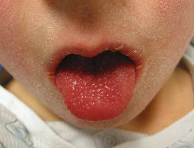
Strawberry tongue Kawasaki disease
Courtesy of Robert Sundel, MD.
- بثورات وزیکولوبولوس:
- آبله مرغان
- ریکتزیال پاکس(پاپول وزیکول اسکار در محل گزش مایت)
- HSV
- Hot tub follicolitis
Hot tub folliculitis
This form of folliculitis is caused by Pseudomonas aeruginosa and can occur after exposure to hot tubs or whirlpools.
Courtesy of Charles V Sanders. The Skin and Infection: A Color Atlas and Text, Sanders CV, Nesbitt LT Jr (Eds), Williams & Wilkins, Baltimore, 1995.
- بثورات ندولر:
- کاندیدیازیس منتشر
- ضایعات منتشر کریپتوکوکی(شبیه مولوسکوم کونتاژیوزوم)
- موکورمیکوزیس
- آسپرژیلوزیس
- سندرم Sweet (ندولها و پلاکهای متعدد ادماتو)
- پتشی و پورپورا:
- مننگوکوکسمی حاد
- اکوویروس(همه نوع ضایعه میدهد)
- کوکساکیویروس(همه نوع ضایعه میدهد)
- VHF (CCHF)
- پلاسمودیوم فالسیپاروم
- RMSF
- سایر موارد:
سرخک آتیپیک، سرخجه ،آدنوویروس،تبدانگ،تب زرد،Variola،EBV،CMV،HBV(پورپورای قابل لمس)
- قارچها فقط نمای ماکولوپاپولار و ندولار میدهند.
- :Rickettsia
- Rickettsii(RMSF) (وکتور: کنه): پتشی
- akari(Rickettsial pox) : وزیکول/بول
- Louse-borne typhos (وکتور: شپش) : پتشی
- Typhi endemic/murine typhus
- Tsutsugamushi (scvub typhus)
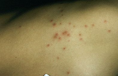
Skin lesions in acute meningococcemia can begin as papules but quickly progress to petechiae and purpura. As seen here, the purpuric lesions can coalesce.
Courtesy of Charles V Sanders. (The Skin and Infection: A Color Atlas and Text, Sanders, CV, Nesbitt, LT Jr (Eds), Williams & Wilkins, Baltimore, 1995).
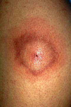
Erythema migrans with central clearing and a necrotic center.
Courtesy of Dori F Zaleznik, MD.
Lymphocutaneous sporotrichosis on the right lower extremity of a young man who overturned his dirt bike and inoculated soil in multiple places in his leg. Following a plastic surgery procedure before the diagnosis of sporotrichosis, new lesions developed at the edge of the skin graft and extended up into the thigh.
Courtesy of Carol A. Kauffman, MD.
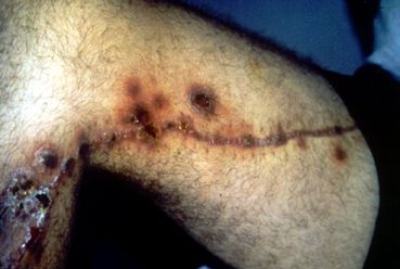
The primary inoculation site in this patient with ulceroglandular tularemia is an ulcerated nodule that is accompanied by lymphadenopathy.
Courtesy of Charles V Sanders. (The Skin and Infection: A Color Atlas and Text, Sanders CV, Nesbitt LT Jr (Eds), Williams & Wilkins, Baltimore, 1995.
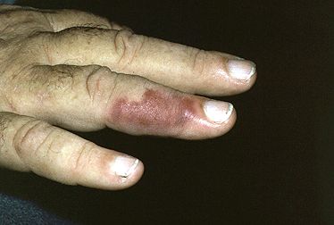
Erysipeloid is the localized cutaneous form of the infection caused by Erysipelothrix rhusiopathiae. In this patient, violaceous maculopapular lesions of the fingers developed after cleaning fish.
Courtesy of Lee T Nesbitt, Jr. The Skin and Infection: A Color Atlas and Text, Sanders CV, Nesbitt LT Jr (Eds), Williams & Wilkins, Baltimore, 1995.
علائم کلینیکی در RMSF:
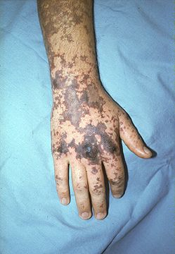
در 3 روز نخست علائم غیر اختصاصی مثل:تب،سردرد،میالژی،تهوع و استفراغ و بیاشتهایی بروز مینماید و سپس ماکولهای محو شونده ابتدا در مچ دست و پا (centripetal) ایجاد میشود که بعد از مدتی هموراژیک شده تبدیل به پتشی میشود.
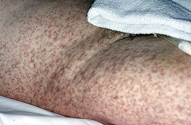
Extensive maculopapular and petechial lesions on the trunk and thigh in a patient with Rocky Mountain spotted fever. The rash characteristically begins on the wrists and ankles and spreads centripetally.
Courtesy of Harold G Muchmore. The Skin and Infection: A Color Atlas and Text, Sanders CV, Nesbitt LT Jr (Eds), Williams &Wilkins, Baltimore, 1995.
درمان:
Doxicyclin 100 mg/po/Iv/bid ×7d or 2 d after afebrile.
(Alt. In pregnancy :Chloramphenicol 50mg/kg/day in 4 div. doses.)
جدول خلاصه ای از انواع بثورات جلدی
|
time
|
Smear+ |
Common pathogens | lesion |
| 12-36 | – | Capnocytophaga conimorsus g- nonninfection | Symmetrical peripheral gangrene, puroura fulminaus, acrocyanosis |
| 12-36 | +
Except RMSF 1-7 d |
N.meningitidis
C.conimorsus Rickettsia |
Multiple purpuric lesions in severely ill patients |
| Severd days | + | Pseudomomas
g- vibrio vulnificur |
Ecthyma gangrenosum, other bullous lesions |
| Severd days | – | Candida, C.neoformans
H.capsulatum, Fusarium |
Macronodular lesions |
| 3-10 d | Occasionally (few) | Neisseria gonorrhoes
Neisseria meningitides (chronic) |
Delayed-unset rash with nonsymmetrical, scattered maculopapular or vesicular lesions |
| 5-10 d | – | Salmonella | Rose spots |
| At presenta | – | SSSS | Toxic erythema |
- Capnocytophaga in splenectomized pts may cause local eschar, sepsis with DIC.